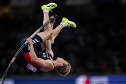
 Sondre Guttormsen


Use Aomi or a guided session to practice what you are reading.

When you vividly imagine throwing a javelin, your brain does not distinguish it from actually throwing one — at least not at the level of motor planning. The premotor cortex, supplementary motor area, basal ganglia, and cerebellum all activate during vivid motor imagery, mirroring the neural architecture of physical execution. This is not a metaphor or a motivational claim. It is a replicable finding from functional neuroimaging, documented across dozens of studies and multiple brain imaging modalities.
Understanding this neuroscience transforms visualization from a feel-good heuristic into a principled training method with predictable physiological effects.
Functional Equivalence: The Central Principle
In 1994, Marc Jeannerod published a landmark paper in Behavioral and Brain Sciences titled "The Representing Brain," which formalized the functional equivalence hypothesis: motor imagery and motor execution are mediated by overlapping neural mechanisms. Using positron emission tomography (PET) and later confirmed with functional magnetic resonance imaging (fMRI), Jeannerod demonstrated that imagined movements activate the same cortical and subcortical structures as executed movements — with the critical exception of the primary motor cortex (M1), which is partially inhibited during imagery to prevent actual movement output.
Subsequent research has refined this picture substantially:
- Lotze and Halsband (2006) reviewed neuroimaging studies across multiple modalities and confirmed that motor imagery consistently activates the premotor cortex, SMA, posterior parietal cortex, basal ganglia, and cerebellum — the same circuit that plans, coordinates, and fine-tunes skilled movement.
- Decety and Grèzes (1999) showed that the degree of neural overlap between imagery and execution depends on the vividness and kinesthetic quality of the imagery, not merely whether the person is "trying to imagine."
- Hétu et al. (2013), in a comprehensive meta-analysis of 122 fMRI studies, mapped the precise anatomical regions activated during motor imagery across upper limb, lower limb, and whole-body tasks, confirming the somatotopic organization of imagined movement (i.e., imagining leg movements activates leg-related motor areas).
The implication for athletes is direct: high-quality motor imagery is not a psychological trick. It is a neural training stimulus that engages the motor planning system through the same circuitry used during physical practice.
What Happens in the Brain During Visualization
Here is a systems-level overview of the neural processes during structured motor imagery:
Stage 1: Movement Planning (Prefrontal Cortex)
The brain begins by constructing the motor plan — the sequence, timing, and force parameters of the intended movement. This engages the dorsolateral prefrontal cortex (DLPFC), the same executive region responsible for strategic decision-making and working memory during competition. The DLPFC integrates contextual information (competition scenario, tactical goals) with stored motor programs to generate a task-appropriate execution plan (Fuster, 2001).
Stage 2: Motor Preparation (Premotor Cortex and SMA)
The premotor cortex and supplementary motor area prepare the motor system for execution. During physical movement, this preparation culminates in a "go" signal to the primary motor cortex. During imagery, the preparation proceeds identically — but the final execution signal is attenuated.
Critically, this preparation is not silent at the muscular level. Electromyographic (EMG) studies have demonstrated that muscles relevant to the imagined task exhibit measurable electrical activity during imagery — a phenomenon termed subliminal motor irradiation (Guillot et al., 2007). The signal is insufficient to produce overt movement, but its presence confirms that the corticospinal pathway is partially engaged. This subliminal activation is believed to contribute to the motor learning effects of mental practice.
Stage 3: Temporal Coordination (Cerebellum)
The cerebellum calibrates the timing and sequencing of multi-joint movements. Its activation during motor imagery explains why temporally accurate imagery (visualizing at real-time speed) produces larger training effects than slow-motion or accelerated imagery (Ivry & Spencer, 2004). The cerebellum is training the same timing parameters during imagery that it coordinates during execution.
Stage 4: Emotional Priming (Limbic System)
When imagery incorporates emotional content — the pressure of a match point, the crowd noise, the adrenaline of a final attempt — the amygdala and associated limbic structures engage. The amygdala modulates the consolidation of motor memories through its influence on long-term potentiation (McGaugh, 2004). This means emotionally charged imagery produces more durable motor traces than emotionally neutral imagery — a finding with direct implications for competition preparation.
Stage 5: Inhibitory Gating (Primary Motor Cortex)
The primary motor cortex (M1) is partially inhibited during imagery, preventing the motor commands from reaching the muscles at full amplitude. This inhibition is mediated by GABAergic interneurons and descending inhibitory signals from prefrontal areas (Jeannerod, 2001). The system effectively "runs the simulation" while blocking the output — analogous to a flight simulator that trains piloting skills without leaving the ground.
Quantifying the Effect: What the Meta-Analyses Show
The performance effects of mental practice have been quantified across multiple meta-analyses:
Driskell, Copper, and Moran (1994) synthesized 35 studies across athletic, musical, and surgical domains. They reported a weighted mean effect size of d = 0.527, representing a meaningful improvement when mental practice is added to physical training. The analysis found three critical moderators:
- Task type: Cognitive tasks (sequence planning, strategy) showed larger effects than purely motor tasks, but motor tasks still showed significant improvement.
- Imagery quality: Structured, vivid imagery produced substantially larger effects than vague or passive imagery.
- Retention interval: Benefits persisted for weeks after the intervention ended, suggesting genuine motor learning rather than transient priming.
Feltz and Landers (1983), in an earlier meta-analysis of 60 studies, reported an average effect size of d = 0.48 for mental practice, and found that the combination of mental and physical practice consistently outperformed physical practice alone.
Schuster et al. (2011) reviewed mental practice effects specifically in rehabilitation contexts, finding that motor imagery accelerated recovery of movement function after stroke and orthopedic surgery — extending the functional equivalence principle beyond sport into clinical neurorehabilitation.
Why Most Athletes Visualize Incorrectly
Despite the robust evidence base, many athletes who attempt visualization report minimal benefit. The research literature identifies several consistent quality failures:
Outcome-focused imagery. Imagining "winning" or "doing well" does not activate specific motor pathways. The brain's somatotopic motor maps require detailed, effector-specific movement imagery to produce neural adaptation. Imagining the feeling of victory is emotionally pleasant but motorically inert.
Postural incongruence. Lying in bed imagining a tennis serve produces weaker corticospinal excitability than sitting upright or standing (Guillot et al., 2007). Physical posture modulates the motor system's receptivity to imagery-based signals — a finding that motivated the "Physical" component of the PETTLEP model (Holmes & Collins, 2001).
Absent emotional engagement. Calm, detached visualization fails to engage the limbic system, forfeiting the memory consolidation benefits of emotional arousal. Athletes who visualize in an analytical mode are training cognitive understanding, not motor preparation.
Temporal distortion. Freezing on a single moment (the finish line, the winning shot) rather than running through the complete performance sequence prevents the cerebellum from rehearsing the timing and coordination patterns needed for execution.
Representational staleness. Using the same visualization script for months, even as technique evolves, creates a mismatch between the mental model and the athlete's current motor capability. The imagery must update with the athlete's development — the "Learning" component of PETTLEP.
Applications Across Athletic Contexts
Pre-Competition Preparation
Spend 10–15 minutes visualizing the performance plan: the specific skills to be executed, in the order they will occur, at real-time speed. Incorporate the competition environment, expected emotional states, and technical priorities. This primes the motor system for the specific demands of the upcoming performance.
Post-Practice Consolidation
After a successful repetition in training, close your eyes and mentally replay it. This doubles the neural reinforcement of that successful movement pattern. Debriefing through imagery — rather than simply moving to the next repetition — has been shown to enhance motor learning rates (Malouin et al., 2013).
Injury Rehabilitation
Research by Cupal and Brewer (2001) demonstrated that athletes who incorporated motor imagery into ACL rehabilitation returned to sport faster and reported lower reinjury anxiety than a standard rehabilitation-only control group. Maddison et al. (2012) found similar results for post-surgical knee rehabilitation, with imagery groups showing greater knee strength and self-efficacy at 6 months post-surgery.
When the body cannot train, the motor planning system can still be exercised. This is one of the most clinically significant applications of the functional equivalence principle.
Rest Days and Detraining Prevention
Motor programs — the neural representations of skilled movements — degrade during extended periods without activation (Wulf & Shea, 2002). Visualization on rest days maintains these representations, preventing the gradual detraining that occurs when the motor system goes unstimulated. It is not a replacement for physical practice, but a neural maintenance strategy that preserves training adaptations between sessions.
Implications for Technology-Delivered Mental Training
The neuroscience of motor imagery provides a clear blueprint for what visualization tools must do to be effective: they must produce imagery that is specific, temporally accurate, emotionally engaged, physically congruent, and environmentally anchored. Generic meditation apps and motivational affirmation generators do not meet these criteria.
At Athlete Mindset, we designed the AI system around these neuroscientific requirements. The app generates sport-specific, PETTLEP-based visualization sessions that guide athletes through detailed, step-by-step motor imagery — matching the structural characteristics that the research identifies as necessary for genuine neural adaptation.
The athletes who dismiss visualization as "woo-woo" have typically only experienced the unstructured variant. The athletes who integrate it as a core training tool have learned that, at the level of the motor cortex, the brain treats high-quality imagery as practice.
Sondre Guttormsen is a two-time Olympian (Tokyo 2020, Paris 2024), Norwegian pole vault record holder (6.06m indoor), 3x NCAA Champion, and Princeton psychology graduate. He founded Athlete Mindset to make evidence-based mental training accessible to every athlete.
References
- Cupal, D. D., & Brewer, B. W. (2001). Effects of relaxation and guided imagery on knee strength, reinjury anxiety, and pain following anterior cruciate ligament reconstruction. Rehabilitation Psychology, 46(1), 28–43.
- Decety, J., & Grèzes, J. (1999). Neural mechanisms subserving the perception of human actions. Trends in Cognitive Sciences, 3(5), 172–178.
- Driskell, J. E., Copper, C., & Moran, A. (1994). Does mental practice enhance performance? Journal of Applied Psychology, 79(4), 481–492.
- Feltz, D. L., & Landers, D. M. (1983). The effects of mental practice on motor skill learning and performance: A meta-analysis. Journal of Sport Psychology, 5(1), 25–57.
- Fuster, J. M. (2001). The prefrontal cortex — an update: Time is of the essence. Neuron, 30(2), 319–333.
- Guillot, A., Lebon, F., Rouffet, D., Champely, S., Doyon, J., & Collet, C. (2007). Muscular responses during motor imagery as a function of muscle contraction types. International Journal of Psychophysiology, 66(1), 18–27.
- Hétu, S., Grégoire, M., Saimpont, A., Coll, M.-P., Eugène, F., Michon, P.-E., & Jackson, P. L. (2013). The neural network of motor imagery: An ALE meta-analysis. Neuroscience & Biobehavioral Reviews, 37(5), 930–949.
- Holmes, P. S., & Collins, D. J. (2001). The PETTLEP approach to motor imagery. Journal of Applied Sport Psychology, 13(1), 60–83.
- Ivry, R. B., & Spencer, R. M. C. (2004). The neural representation of time. Current Opinion in Neurobiology, 14(2), 225–232.
- Jeannerod, M. (1994). The representing brain: Neural correlates of motor intention and imagery. Behavioral and Brain Sciences, 17(2), 187–202.
- Jeannerod, M. (2001). Neural simulation of action: A unifying mechanism for motor cognition. NeuroImage, 14(1), S103–S109.
- Lotze, M., & Halsband, U. (2006). Motor imagery. Journal of Physiology–Paris, 99(4–6), 386–395.
- Maddison, R., Prapavessis, H., Clatworthy, M., Hall, C., Foley, L., Harper, T., ... & Brewer, B. (2012). Guided imagery to improve functional outcomes post-anterior cruciate ligament repair: Randomized-controlled pilot trial. Scandinavian Journal of Medicine & Science in Sports, 22(6), 816–821.
- Malouin, F., Jackson, P. L., & Richards, C. L. (2013). Towards the integration of mental practice in rehabilitation programs: A critical review. Frontiers in Human Neuroscience, 7, 576.
- McGaugh, J. L. (2004). The amygdala modulates the consolidation of memories of emotionally arousing experiences. Annual Review of Neuroscience, 27, 1–28.
- Schuster, C., Hilfiker, R., Amft, O., Scheidhauer, A., Andrews, B., Butler, J., ... & Ettlin, T. (2011). Best practice for motor imagery: A systematic literature review on motor imagery training elements in five different disciplines. BMC Medicine, 9, 75.
- Wulf, G., & Shea, C. H. (2002). Principles derived from the study of simple skills do not generalize to complex skill learning. Psychonomic Bulletin & Review, 9(2), 185–211.
One useful idea. One repeatable rep.
Build a mental routine you can return to when the moment gets loud.